
Figure 1.
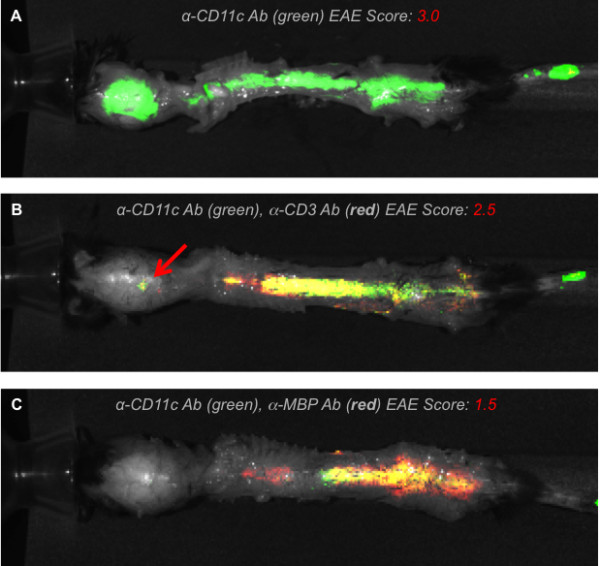
Distribution of CD11c+ dendritic cells in d16 experimental autoimmune encephalomyelitis mice. Ex vivo near-infrared fluorescence imaging of d16 experimental autoimmune encephalomyelitis (EAE) mice showing the distribution of CD11c+ dendritic cells (DCs) in the context of CD3+ T cells and myelin basic protein (MBP). (A) Anti-CD11c antibody only (green) signal from DCs in a mouse with severe degree of EAE. (B) Mouse with moderate EAE score shows signal from both CD11c+ DCs (green) and CD3+ T cells (red) along the thoracic and lumbar spine. (C) Mouse exhibiting mild EAE shows a high degree of co-localization between CD11c+ DCs and MBP signal.