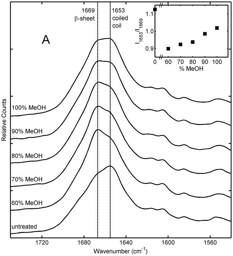
Figure 1. Raman spectra of honeybee silk sponges immersed for 48 hours in aqueous methanol solutions containing 60–100% methanol.
Inset shows the approximate ratio of Raman intensity at 1653 cm−1 (attributed to coiled coil protein structure) to intensity at 1669 cm−1 (attributed to ß-sheet protein structure) for different samples.

