Figure 1.
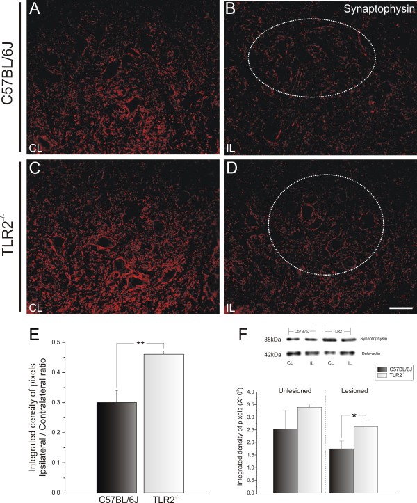
Representative images of synaptophysin immunostaining in C57BL/6J wild-type (WT) and Toll-like receptor (TLR)2 knockout (KO) mice (TLR2−/−) 1 week after unilateral axotomy. Note that 1 week after lesioning, there was a strong decrease in labeling, especially in the areas surrounding the motor neurons. This decrease was more intense in (B) C57BL/6J mice than (D) in TLR2−/− mice. (A,C) Contralateral side of C57BL/6J WT and TLR2−/− mice, respectively. The dashed circle indicates the motor nucleus containing the alpha motor neurons. (E) Graph representing the ipsilateral:contralateral (IL:CL) ratio of the integrated density of pixels ** P<0.01. (F) Western blot analysis of synaptophysin expression in WT and TLR2 KO mice. Note the significant preservation of synaptophysin in TLR2−/− after peripheral axotomy. β-Actin was used as sample loading control. * P<0.05. Scale bar: 50 μm.

