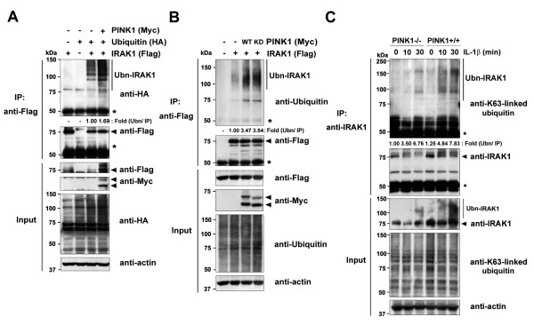
Figure 5.
PINK1 increases Lys63-dependent polyubiquitination of IRAK1. (A) HEK293 cells were transfected for 48 hours with HA-ubiquitin, Flag-IRAK1, or Myc-PINK1, alone or in combination. Cell lysates were immunoprecipitated with the Flag antibody, followed by immunoblotting with the HA antibody. Expression of transiently transfected proteins was confirmed by immunoblotting with the HA, Flag, or c-Myc antibodies. *IgG heavy chains.Actin served as a loading control. (B) 293 IL-1RI cells were transfected for 42 hours with Flag-IRAK1 alone or together with either Myc-tagged wild-type PINK1 (WT) or its kinase-defective mutant (KD), and treated for 15 minutes with 10 ng/ml IL-1β. Cell lysates were immunoprecipitated with the Flag antibody, followed by immunoblotting with the ubiquitin antibody. (C) PINK1−/− and PINK1+/+mouse embryonic fibroblasts were treated with 50 ng/ml IL-1β for the indicated times. Cell lysates were immunoprecipitated with the IRAK1 antibody, and immunoblotted with the Lys63-specific ubiquitin antibody. The relative polyubiquitinated IRAK1 levels were quantified and denoted below the upper panel:(A), (B) (ubiquitinated IRAK1/IRAK1 input)/IRAK1 IP, or (C) (endogenous ubiquitinated IRAK1/endogenous IRAK1)/endogenous IRAK1 IP.IRAK1, IL-1 receptor-associated kinase 1; PINK1, PTEN-induced putative kinase 1.

