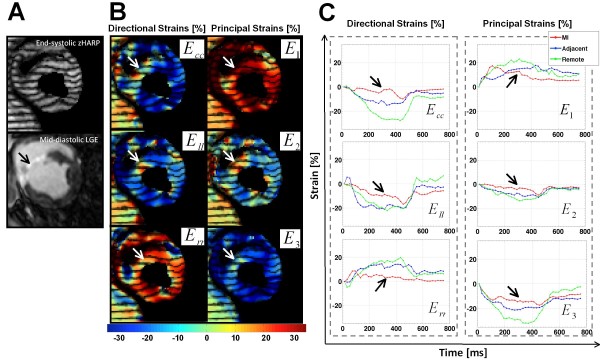
Figure 4.
Example mid-ventricular slice at early post-MI. (A) End-systolic zHARP tagging image (top) with paired mid-diastolic LGE image (bottom). (B) End-systolic directional and principal strains overlaid on the zHARP image. Variation of the strain in infarct region from the remote region is consistent with enhancement in LGE image (white arrows). (C) Average directional strains (Ecc = circumferential strain, Ell = longitudinal strain, Err = radial strain), and principal strains (E1, E2, E3) are displayed in the infarct, adjacent, and remote segments in one cardiac cycle.

