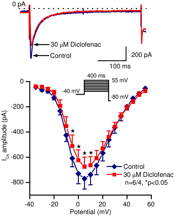
Figure 6. Effect of diclofenac on the L-type calcium current in canine ventricular myocytes.
Top panel shows representative current traces, bottom panel represents current – voltage relationships under control conditions and in the presence of 30 µM diclofenac. Inset indicates the voltage protocol applied during measurements. Data are expressed as mean ± SEM, n = number of measurements/number of animals.

