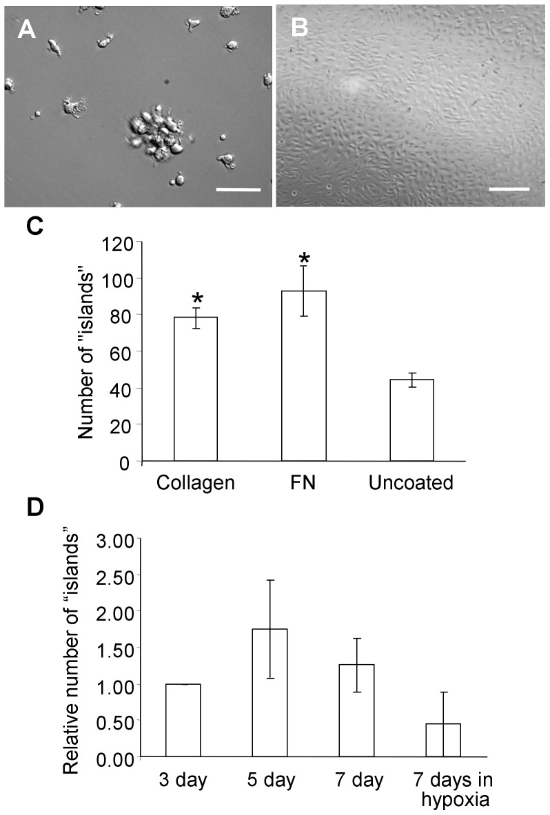
Figure 1. Phenotype of cells arising from peripheral blood mononuclear cells.
Phase contrast images of cell “islands” (A) and an endothelial clone (B). Scale bar = 100 µm (C) Effect of different plating conditions on colony “island” formation of bovine mononuclear cells – the number of colony “islands” counted on collagen and fibronectin (FN) coated plates was statistically greater (*p<0.05) than from non-coated (control) plates. (D) The number of colony “islands” observed in non-coated plates where non-adherent cells were removed 3, 5 or 7 days after incubation in normoxic conditions and 7 days after incubation in hypoxic conditions. No statistically significant difference (p>0.05) was observed.

