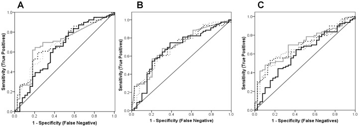
Figure 4. AUROC for IL-6, IL-8 and VEGF.
The lowest area under receiver operating characteristic (AUROC) were determined after protein normalization as represented by the solid black curve which was always closest to the diagonal reference line i.e., IL-6 = 0.634 (0.523 to 0.745); IL-8 = 0.677 (0.570 to 0.784); and VEGF = 0.609 (0.501 to 0.716). The AUROCs for uncorrected biomarker levels (thick grey curve), and those standardized using osmolarity (dashed black curve) or creatinine (dashed grey curve) were very similar for individual biomarkers : (A) IL-6 = 0.693 (0.592 to 0.794), 0.683 (0.582 to 0.784) and 0.678 (0.578 to 0.779), respectively; (B) IL-8 = 0.706 (0.608 to 0.804), 0.701 (0.603 to 0.799) and 0.694 (0.592 to 0.795), respectively; and (C) VEGF = 0.705 (0.610 to 0.799), 0.687 (0.591 to 0.783) and 0.680 (0.583 to 0.777), respectively. Figures in brackets are 95% Confidence Intervals.

