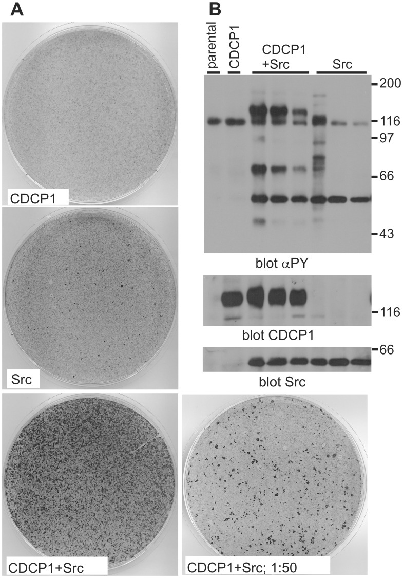
Figure 1. Co-overexpression of Src and CDCP1 leads to focus formation.
(A) Fifty thousand NIH3T3 cells were infected with 500.000 retroviruses encoding CDCP1 or Src individually or together. After 48 h the cells were reseeded in a 10 cm-dish and grown for 3 weeks, with 3 media changes per week. In parallel, a 2% aliquot was taken from the CDCP1/Src infected cells and reseeded together with 150.000 parental NIH3T3 cells (1∶50 dilution). Finally, the cells were stained with crystal violett. (B) Parental NIH3T3 cells, cells infected with the CDCP1 encoding retrovirus or individual foci derived from infections at lower m.o.i. than in A that had been isolated and expanded, were lysed, aliquots with similar amounts of protein run on an SDS-PAGE, proteins transferred to nitrocellulose and the filter blotted with the indicated antibodies. αPY, anti-phosphotyrosine. Size markers (kDa) are indicated.

