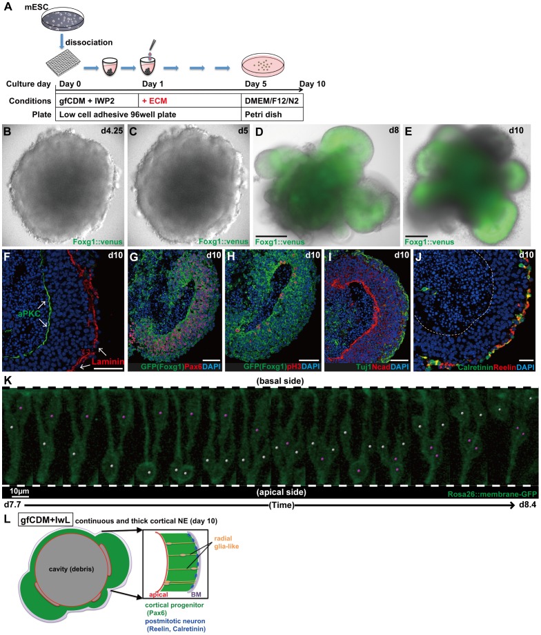
Figure 2. Formation of continuous cortical NE with a clear A–B polarity.
(A) Schematic of SFEBq/gfCDM+IwL culture. (B–C) Live imaging showing the formation of continuous NE in SFEBq/gfCDM+IwL culture on day 4.25 (B) and on day 5 (C). (D–E) Formation of continuous NE expressing Foxg1::venus on day 8 (D) and on day 10 (E). (F–J) Immunostaining of Foxg1::venus+ thick NE on day 10. (F) Immunostaining for the basement membrane marker laminin and the apical marker aPKC. (G) Cortical progenitors (Foxg1::venus+/Pax6+) occupied the majority of Foxg1::venus+ NE. (H) Mitotic cells (pH3+) located on the apical surface. (I) Postmitotic neurons (Tuj1+) in the superficial-most layer. (J) Immunostaining for Calretinin and Reelin. (K) Live imaging for interkinetic nuclear migration in continuous cortical NE generated from mESCs. Partial labeling was done by mixing Rosa26::membrane-GFP ESCs with non-labeled parental EB5 cells. Snapshots of two neural progenitors (days 7.7–8.4; also shown in Movie S2) undergoing interkinetic nuclear migrations (marked with white or pink dots). (L) Schematic of the formation of continuous cortical NE under gfCDM+IwL conditions on day 10. A dashed line indicates the apical border of NE. Scale bars, 200 µm (D–E); 50 µm (F–J); 10 µm (K).

