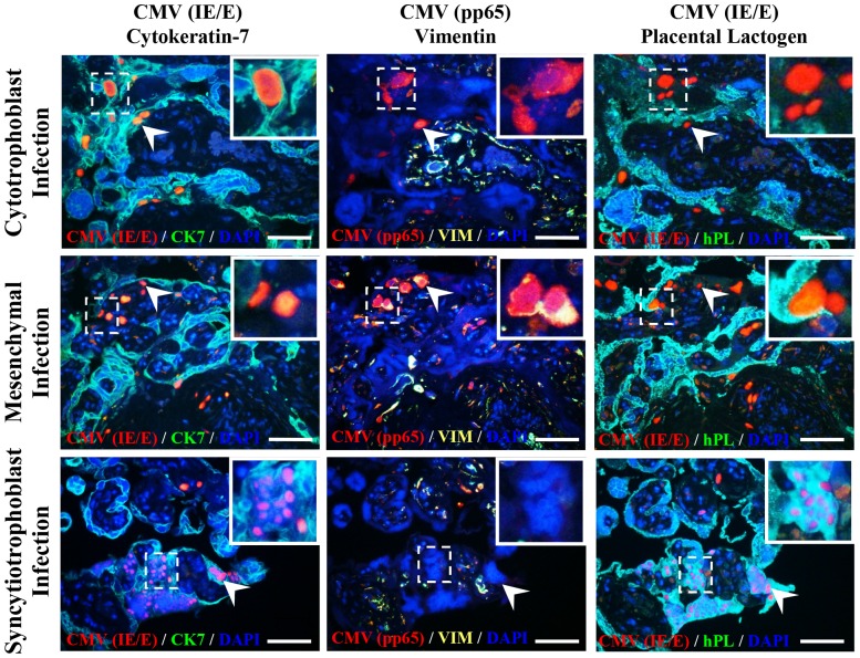
Figure 3. CMV actively replicates in cytotrophoblast and mesenchymal cells, but not syncytiotrophoblasts, of placental villous explants.
CMV laboratory strain AD169 and wild-type Merlin infected syncytiotrophoblasts (CK7+/VIM−/hPL+), cytotrophoblasts (CK7+/VIM−/hPL−) and mesenchymal cells of the villous stroma (CK7−/VIM+/hPL−) as determined by staining for CMV immediate early/early protein. Active replication (CMV IE/E+, pp65+) was observed in both cytotrophoblasts and mesenchymal cells but not syncytiotrophoblasts (CMV IE/E+, pp65−) (inserts and arrow heads). Representative images are of 4 µm consecutive histological sections of AD169 infected villous explants 12 days post inoculation. No difference in cellular tropism was observed between AD169 and Merlin strains. Scale bars represent 100 µm.

