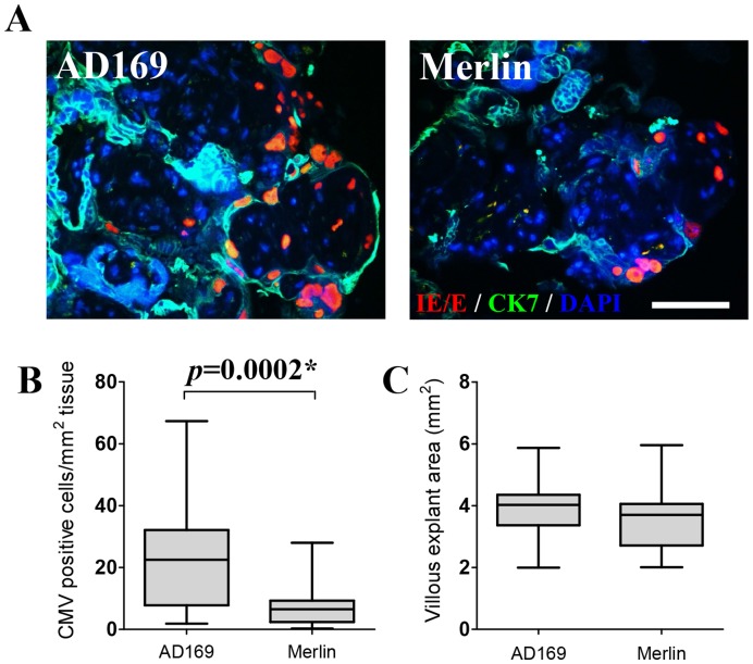
Figure 4. CMV infection is significantly greater in AD169- compared with Merlin-infected placental villous explants.
(A) Representative images of CMV immediate early/early (IE/E) antigen detection in AD169- (left) and Merlin-infected (right) placental villous explants 12 days post inoculation (CK7; Cytokeratin-7). Scale bar represents 70 µm. (B) Number of cells per mm2 of villous explant tissue expressing CMV IE/E protein in AD169- compared with Merlin-infected explants. (C) No significant differences in villous explant area (mm2) were observed between the CMV-infected explant groups (p = 0.1). Data presented as box plots with median value, Q1, Q3 and range. Significant differences between groups (denoted as *) were determined using a one-tailed Spearman’s correlation.

