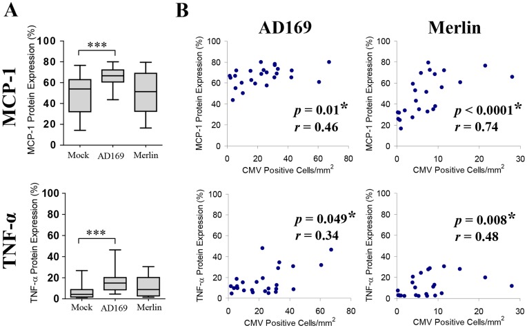
Figure 5. CMV infection of placental villous explants results in upregulation of MCP-1 and TNF-α expression.
(A) MCP-1 and TNF-α expression in mock-infected, AD169-infected (0.2 pfu/ml) and Merlin-infected (0.02 pfu/ml) villous explants. Data presented as box plots with median value, Q1, Q3 and range. Significant differences between groups were determined with the Mann-Whitney U test and threshold for significance adjusted to account for multiple comparisons using Bonferroni’s correction; *p<0.025, **p<0.005 and ***p<0.0005. (B) Correlation between degree of CMV placental explant infection with corresponding MCP-1 and TNF-α expression. MCP-1 and TNF-α protein expression was plotted against the number of cells expressing CMV immediate early/early (IE/E) protein per mm2 of AD169 and Merlin infected villous explant tissue. Significant correlations (denoted by *) were determined by two-tailed nonparametric spearmans correlation with trend plotted as straight lines.

