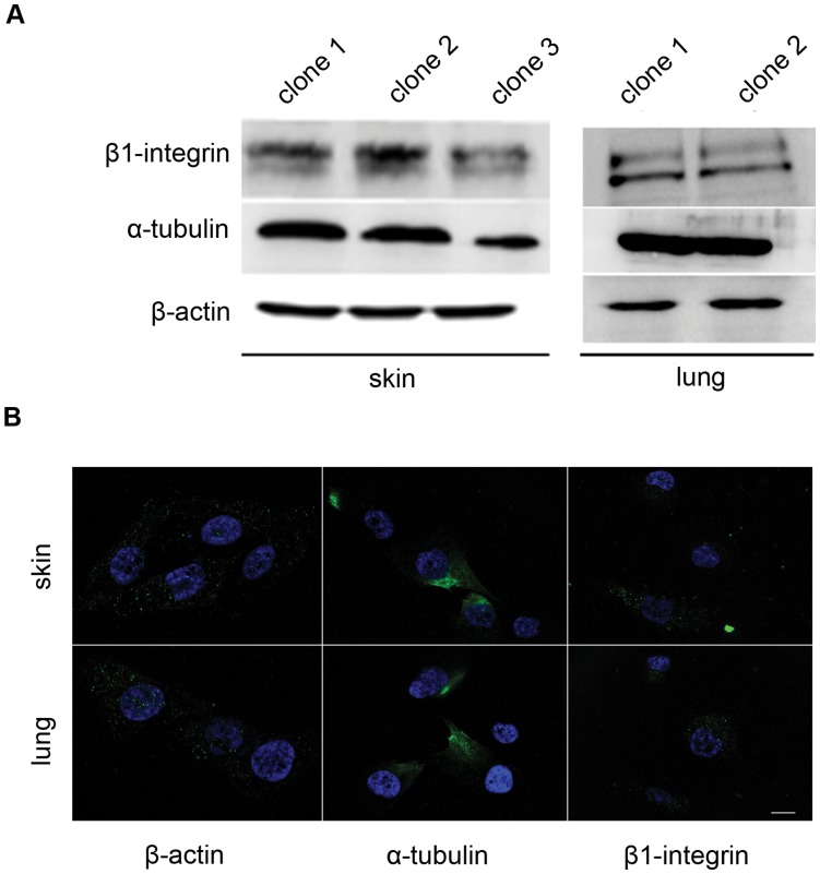
Figure 3. Cytoskeletal protein expression and localization in MPCs.
A. Western blot showing constitutive expression of cytoskeletal components in mesenchymal precursors from skin and lung. One out of three independent experiments is shown. B. Representative fluorescence images of skin and lung MPCs, showing the distribution of α-actin, β-tubulin and β1-integrin rabbit anti-mouse antibody). Scale bar, 1 µM.

