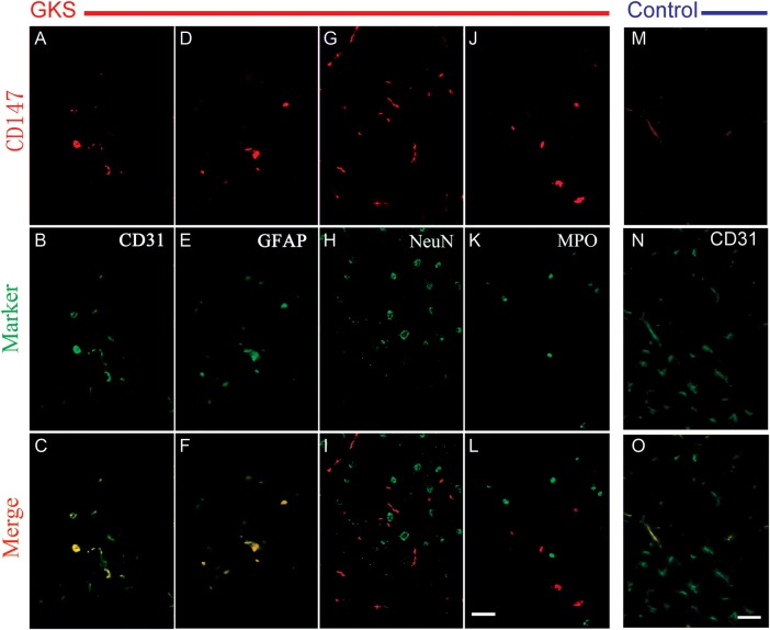
Fig. 3.
Representative photomicrographs of double fluorescent staining of CD147 with different cell markers in the irradiated brain tissue.
Brain sections were stained for CD147 (red), CD31 (green), GFAP (green), and MPO (green). Co-localization of red and green fluorescence is shown in yellow. CD147+ cells are colocalized with CD31+ endothelial cells and GFAP+ astrocytes in the irradiated tissue. CD147+ cells are only colocalized with CD31+ endothelial cells in the control tissue. Scale bar: 50 µm.

