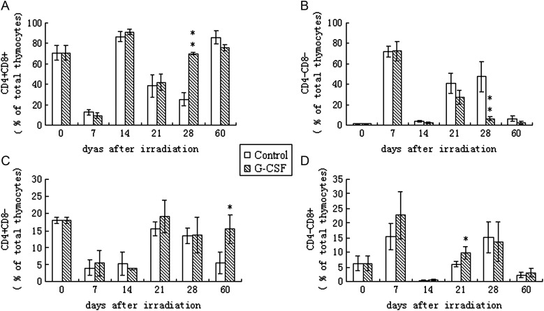
Fig. 2.
Effects of G-CSF administration on the thymocyte subset distribution.
BALB/c mice were exposed to 6 Gy of total body irradiation and were treated with 100 μg/kg G-CSF or an identical volume of PBS (control) once daily for 14 days. Thymocyte subsets CD4 + CD8+ (Fig. 2A), CD4–CD8– (Fig. 2B), CD4 + CD8– (Fig. 2C) and CD4–CD8+ (Fig. 2D) were detected once a week post-irradiation by flow cytometry. Values represent the mean ± SD of six independent animals per group. *P < 0.05, **P < 0.01 compared with PBS-treated control.

