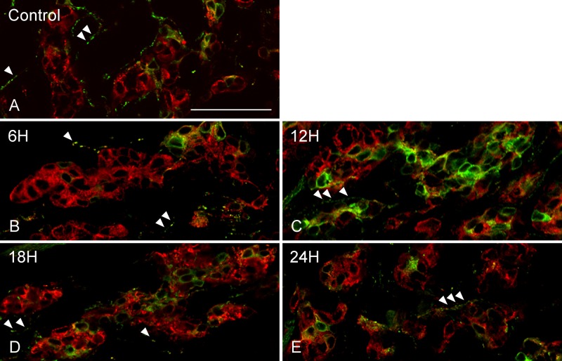
Figure 3.
Double immunofluorescence images of dopamine β-hydroxylase (DBH; green) and synaptophysin (red). Immunoreactivity for DBH was observed in the cytoplasm of glomus cells immunostained for synaptophysin. A few DBH-immunopositive glomus cells were scattered in the carotid body of controls (A) and rats exposed to hypoxia for 6 hr (B). In rats exposed to hypoxia for 12 hr, the number and fluorescent intensity of DBH-immunoreactive glomus cells were increased (C). DBH-immunopositive glomus cells were lower in number in rats exposed to hypoxia for 18 hr (D) and 24 hr (E) than that of rats exposed to hypoxia for 12 hr. DBH immunoreactivity was also observed in varicose nerve fibers (A–E, arrowheads), and there were no obvious differences in DBH-immunoreactive nerve fibers between experimental groups (A–E). Scale bars = 50 µm.

