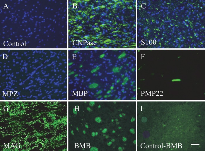
Figure 1.
Expression of constituent myelin proteins and BMB staining in serial sections of brain striatum. The control, containing the secondary antibody only (no primary antibody), is shown in A. Staining with an antibody against CNPase (B), S100 (C), MPZ (D), MBP (E), PMP22 (F), and MAG (G) is shown. BMB staining is shown in (H) along with its control (I), a tissue section with no BMB imaged under the same conditions as with BMB. Nuclear staining by DAPI is also shown in panels A to E. Bar = 50 µm.

