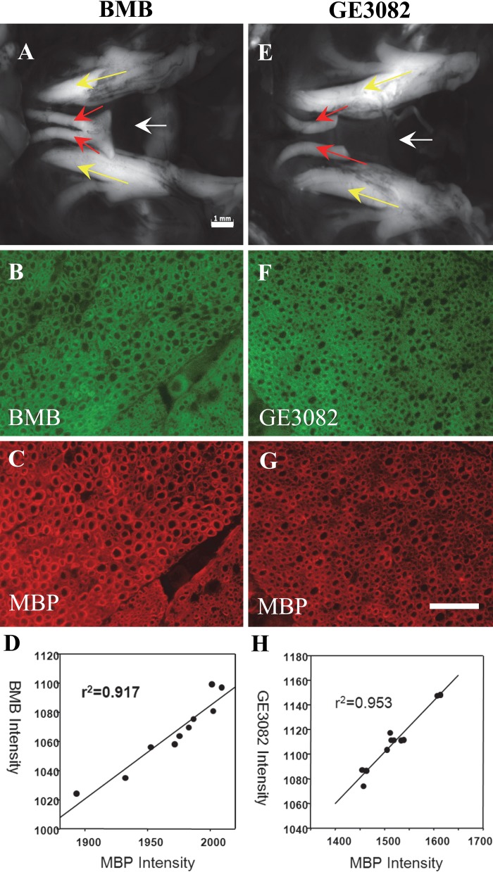
Figure 5.
BMB and GE3082 were injected intravenously in mice and the nerves were imaged. In vivo fluorescence images of the trigeminal nerves of mice treated with BMB (A) and GE3082 (E). Yellow arrow = trigeminal nerves; red arrow = optic nerves; white arrow = sphenoid bone lying under the nerves. The nerves were resected and co-stained with the myelin basic protein (MBP) antibody. BMB and GE3082 staining in the sectioned trigeminal nerve is shown in panels B and F, respectively. MBP staining of BMB- and GE3082-stained sections is shown in panels C and G, respectively. Correlation between MBP and BMB staining is shown in panel D, whereas that of MBP and GE3082 is shown in panel H. Bars: A, E = 1 mm; B–G = 50 µm.

