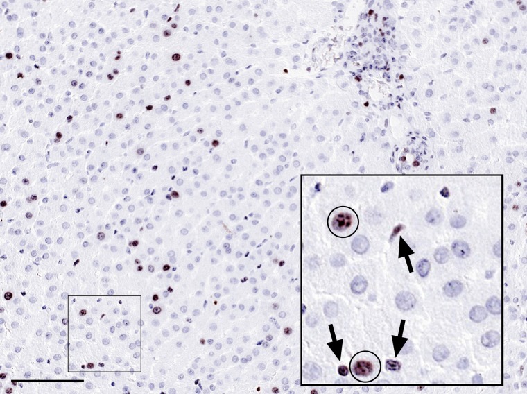
Figure 1.
Formalin-fixed and paraffin-embedded rabbit liver tissue section showing the presence of a substantial number of Ki67-positive nuclei in brown and weakly counterstained with hematoxylin. It is mostly unclear if these cells are proliferating hepatocytes or non-parenchymal cells, of which sinusoidal lymphocytes may be particularly difficult to distinguish from hepatocytes, both having round nuclei. Insert shows a high magnification with Ki67-positive nuclei from hepatocytes (circles), whereas the nuclei depicted by arrows are non-parenchymal cells in the sinusoidal space, including lymphocytes. Bar = 0.1 mm.

