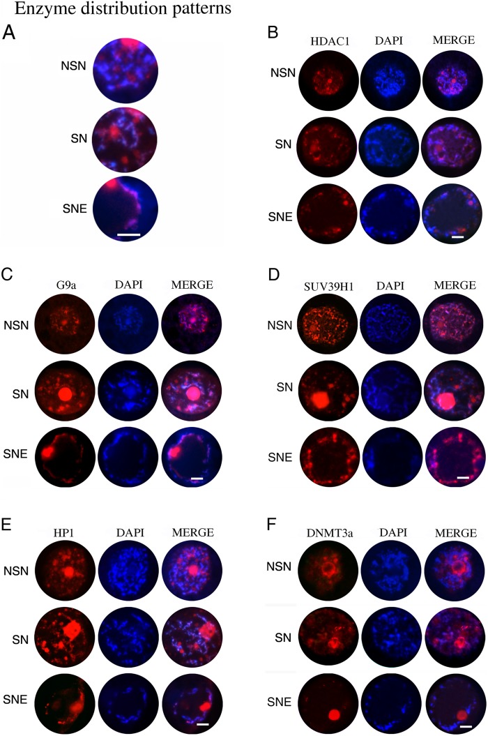
Figure 5.
(A) Summary of all enzyme distribution patterns inside the germinal vesicles (GVs): NSN, a typical GV with a diffuse chromatin (NSN) where the analyzed enzymes show a diffuse and punctiform immunopositivity near the chromatin; SN, a GV example of an early antral follicle with a condensed chromatin around the nucleolus (SN) where the immunofluorescence of the enzymes appeared almost superimposed to the chromatin; and SNE, a GV example of an antral follicle where the chromatin is partially localized around the nucleolus and the nuclear envelope (SNE configuration). There is a clear immunopositivity for the analyzed enzymes that co-localizes perfectly with the chromatin. (B–F) Digital images of sheep GVs showing immunopositivity for HDAC1 (B), G9a (C), SUV39H1 (D), HP1 (E), and Dnmt3a (F). The enzymes are all visible in red (left images), chromatin is in blue (middle images), and merged images (right images) show both. Bar = 100 µm.

