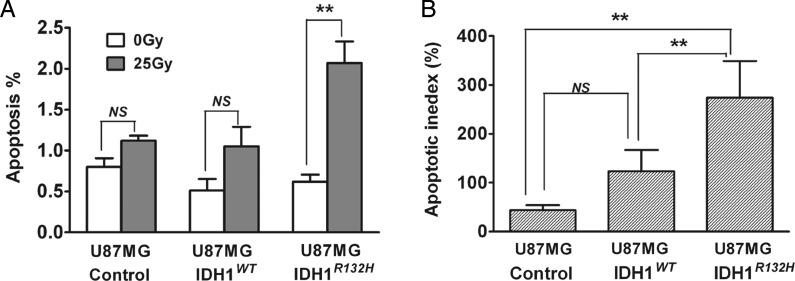
Fig. 4.
U87MG-IDHR132H cells showed significant increase in apoptosis following radiation treatment. (A) Apoptosis was assessed by YO-PRO-1 and propidium iodide (PI) staining after 48 h radiation treatment (0 Gy or 25 Gy). Harvested cells were evaluated by flow cytometry. Column plot summarized 3 independent experiments, each performed with triplicates (mean ± SEM, n =3). **P < .01 compared with U87MG-control and U87MG-IDH1WT cells. (B) Apoptosis % was normalized to U87MG-control cells treated with 0 Gy and expressed as mean apoptotic index (mean ± SEM, n =3). **P< .01 compared with U87MG-control and U87MG-IDH1WT cells.

