
Figure 8.
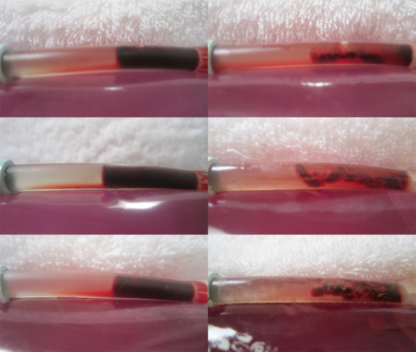
Pre and post LFVP treated samples (SK enriched group). Images (left) depict the pre-condition of three SK enriched LFVP samples (images, top to bottom taken from sample no.’s 6, 11 and 15 respectively). Images (right) depict the samples following 20 minutes of pulsated fluid coordinated with “diastolic” timed LFVP. Note the obvious decrease in clot size and development of substantial clot length fluid channels within the catheter segment.