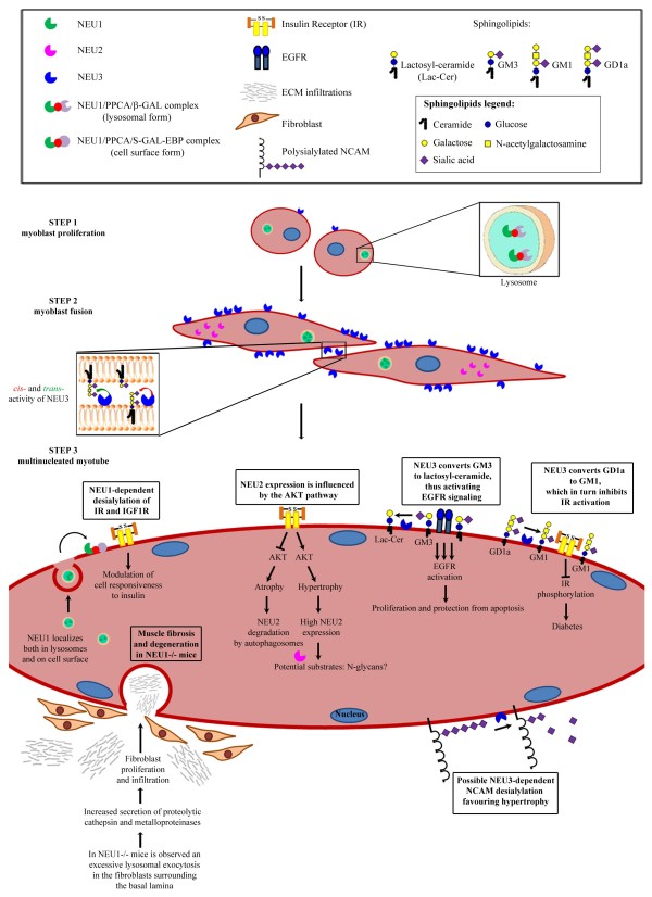
Figure 1.
Cartoon depicting the role of sialidases during the multistep process of myogenesis. STEP 1: During myoblast proliferation, NEU1 is complexed with PPCA and β-GAL and its activity is mainly detectable within lysosomes, whereas plasma membrane-associated NEU3 is present at low levels. On the other hand, the cytosolic NEU2 is absent. STEP 2: Myoblast fusion is characterized by an early and transient increase of NEU1 expression as well as by a long-lasting increment of both NEU2 and NEU3 expression, the latter being involved in cell-cell recognition by working on gangliosides resident on the same cell (cis-activity) or on adjacent cells (trans-activity), as shown in the enlarged box. STEP 3: In differentiated myotubes, NEU1 participates in the degradation of sialo-glycoconjugates in lysosomes but it is also targeted to the cell surface, where it may desialylate IR and IGF1R, thus affecting their responsiveness to insulin. Since NEU1 limits the lysosomal exocytosis in the fibroblasts surrounding the myofibers, NEU1 −/− mice exhibit muscle degeneration due to infiltration of connective tissues. In the cytosol of myotubes, NEU2 expression is modulated mainly through the AKT pathway during hypertrophy and atrophy. In this compartment, cytosolic N-glycans may represent suitable substrates of this enzyme. At the plasma membrane, NEU3 positively affects EGFR signaling by converting GM3 to lactosyl-ceramide, while it blocks the IR activity by conversion of GD1a to GM1. Cell surface sialylated molecules, such as NCAM, may be a target of NEU3 activity. Note that depiction of sugar chains corresponds to the simplified style used in [144]

