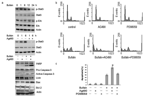
Figure 3.
Bufalin-induced apoptosis was enhanced by inhibition of Jak-STAT3 not ERK. (a) Western blot detected the changes in the protein expression of p-STAT3, STAT3, P-ERK, ERK, PARP, BAX, BCL-2, caspase-3 and livin in SW620 cells that were exposed to 80 nmol/L bufalin alone or 80 nmol/L bufalin plus 20 μmol/L AG490. (b) Cells were exposed to 80 nmol/L bufalin for 24 h in the absence or presence of 20 μmol/L AG490 or 20 mmol/L PD98059. The cell cycle was analyzed by flow cytometry after staining with propidium iodide. The experiments were reproduced three times. One representative experiment is shown. (c) The percentage of apoptotic cells was analyzed by flow cytometry. *P< 0.05 vs. bufalin alone. Data are the mean ± SD of three independent experiments.

