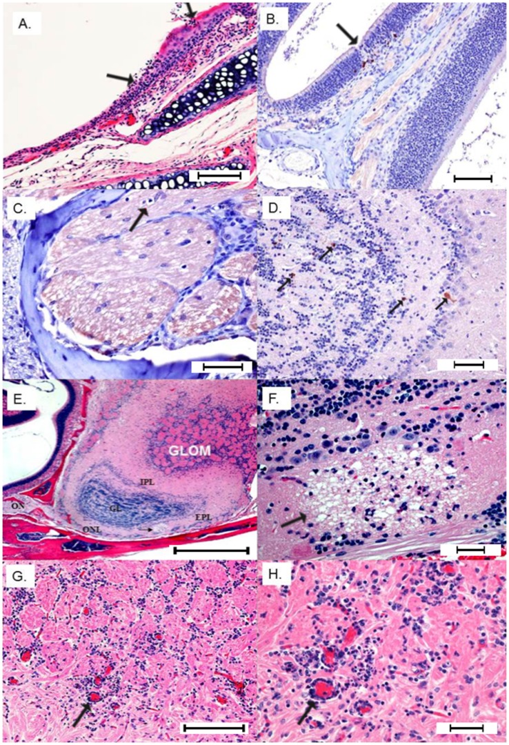
Figure 3. I. Extraneural lesions 24 hpi.
A) Focal erosion/necrosis of the olfactory mucosa with deciliation of the flanking epithelium and neutrophil infiltration into the mucosa and submucosa (Bar = 100 µm). B) IHC positive staining for FLUC is highlighted in a few neuropeithelial cells subjacent to a focal loss of olfactory mucosa (Bar = 200 µm). II- Neuroinvasion from olfactory nerve 48–72 hpi. C) Terminal end of olfactory nerve shows a digestion chamber (arrow) with occasional lymphocytes infiltrating vacuolated branches (Bar = 100 µm). D) Early immunoreactivity (anti-FLUC) in the main olfactory bulb involving scattered neurons in the external plexiform and granular layers (Bar = 100 µm). E) Sagittal section H&E showing the connection between olfactory nerve (ON) and main olfactory bulb layers affected by multifocal necrotizing lesions with associated status spongiosis and infiltration of neutrophils. Glom = glomerular layer; EPI = external plexiform layer; IPL = internal plexiform layer; and ONL = olfactory nerve layer at the ventrum of the olfactory bulb (Bar = 400 µm). F) Neuropil of the olfactory nerve layer shows a large vacuolar lesion (demyelination) with individual neuronal necrosis and infiltration of small numbers of neutrophils (Bar = 100 µm). G) Perivascular cuffs and multifocal gliosis in the glomerular layer 72 hpi (Bar = 200 µm). H) Close-up view of the congested glomerular vessels with pleocellular perivascular cuffs comprising moderate numbers of neutrophils, lymphocytes and glial cells (Bar = 100 µm).

