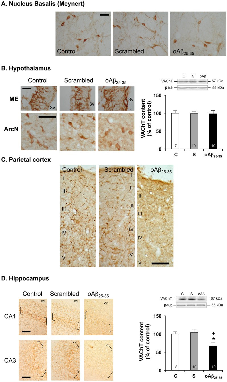
Figure 10. Cholinergic system.
Effects of oAβ25–35 (10 µg/rat) icv injection on VAChT immunolabelling within the nucleus basalis of Meynert (A), mediobasal hypothalamus (B), parietal cortex (C) and hippocampus (D) determined in control untreated rats and 6 weeks after Aβ25–35 injection. In (B): 3v: third ventricle. In (C): levels I to V cortical layers are indicated. In (D): brackets show the hippocampus granular cell layer. cc: corpus callosum. Scale bars = 100 µm. Variations in VAChT levels in the hypothalamus (B) and hippocampus (D), determined in rats by western blot 6 weeks after icv injection of scrambled Aβ25–35 peptide (10 µg/rat; negative control) or oAβ25–35 (10 µg/rat). VAChT (70 kDa) variations were normalized with β-tubulin (β-tub, 55 kDa) variations and compared with untreated rats (control group: C). The results are expressed as means ± SEM. *p<0.05 and **p<0.01 vs. control group, +p<0.05 and ++p<0.01 vs. scrambled treated rats. The number of animals in each group is indicated within the columns.

