
Figure 1. Phospho-Akt expression in mitotic cells in retinal monolayer cultures and intact embryo retina.
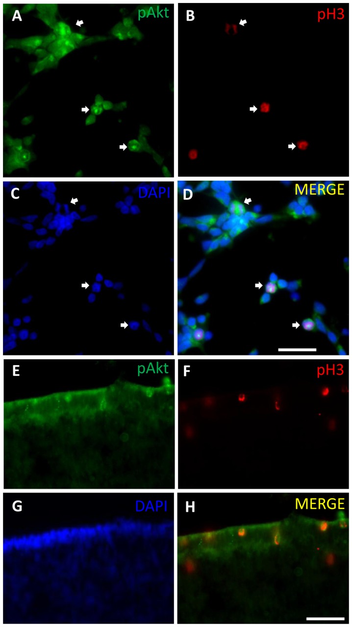
(A–D) Retinal cell cultures from 7-day-old chick embryos maintained for 1 day (E7C1) were fixed and assayed for immunofluorescence against phospho-Akt (A) and phospho-histone H3 (B). Nuclei were stained with DAPI (C). Merged figures in D. (E–H) Retinal sections from 8-day-old embryo retinas were assayed for immunofluorescence against phospho-Akt (E) and phospho-histone H3 (F). DAPI staining of nuclei (G). Merged figures in (H). Arrows point to double labeled mitotic cells. Scale bar = 20 µm in A–D and 30 µm in E–H.