
Figure 4. PI3K inhibition decreases phospho-Akt and phospho-GSK-3 levels in retinal cells.
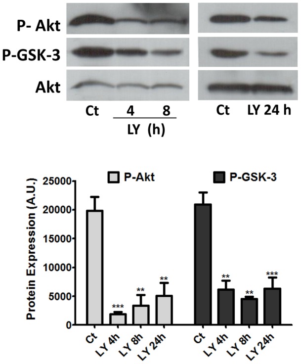
Retinal explants from 7-day-old chick embryos were treated with 25 µM LY 294002 for 4, 8 or 24 h and protein extracts processed for detection of phospho-Akt and phospho-GSK3 expression. Gel loading was assessed with anti-Akt antiserum. Blots were quantified by densitometry and data are expressed as the mean ± S.E.M. of arbitrary units. **p<0.01 and ***p<0.001 relative to control. In both cases, representative blots from at least 3 independent experiments are shown. Ct = control explants cultivated without drug.