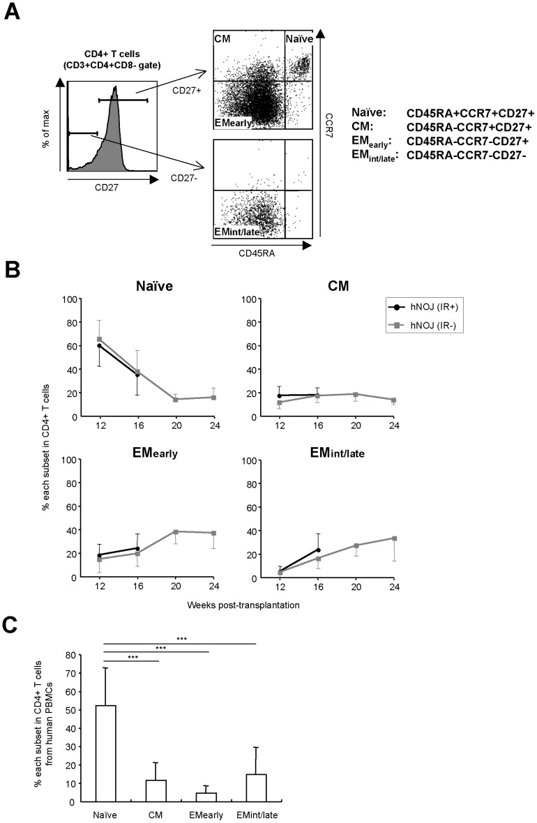
Figure 3. Differentiation of CD4+ T Cells in hNOJ mice.
(A) Naïve, CM, EMearly, and EMint/late subsets of CD4+ T cells (gated on CD3+CD4+CD8−) were defined as CD45RA+CCR7+CD27+, CD45RA−CCR7+CD27+, CD45RA−CCR7−CD27+, and CD45RA−CCR7−CD27−, respectively, by flow cytometry. (B) Changes in the percentage of naïve, CM, EMearly, and EMint/late subsets within the peripheral blood CD4+ T cell populations isolated from hNOJ (IR+) and hNOJ (IR−) mice (n = 18 and n = 6, respectively). Data are expressed as the mean ± SD. (C) Percentage of CM, EMearly, and EMint/late subsets within human peripheral blood CD4+ T cells. Data are expressed as the mean ± SD (n = 10). Significant differences (*** P<0.001) were determined by Tukey’s multiple comparison test.

