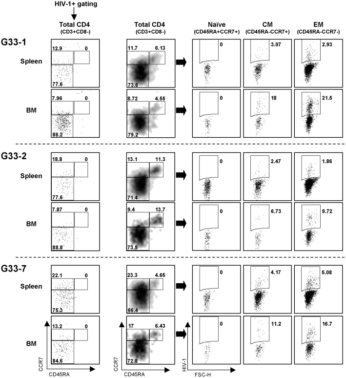
Figure 8. Identification of R5 HIV-1-infected cells in hNOJ mice.
Three hNOJ (IR−) mice (G33-1, G33-2, and G33-7), all at 13 wk post-transplantation, were challenged intravenously with HIV-1NL-AD8-D. At 2 wk post-challenge, the mice were sacrificed and the infected cells in the spleens and BM were analyzed by flow cytometry. Each CD4+ T cell subset (Naïve, CM, or EM) was defined as outlined in the legend to Figure 6. Infected cells were identified based on their expression of the fluorescent reporter, DsRed.

