
Figure 1. PB assembly and disassembly during the cell cycle.
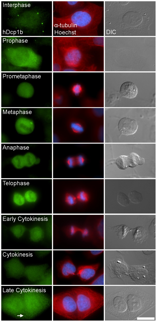
Immunofluoresence staining of endogenous hDcp1b (green), α-tubulin (red), DNA (Hoechst, blue) and DIC images show that PB structures disassemble during cell division. (Bar 20 µm).
Official websites use .gov
A
.gov website belongs to an official
government organization in the United States.
Secure .gov websites use HTTPS
A lock (
) or https:// means you've safely
connected to the .gov website. Share sensitive
information only on official, secure websites.
Immunofluoresence staining of endogenous hDcp1b (green), α-tubulin (red), DNA (Hoechst, blue) and DIC images show that PB structures disassemble during cell division. (Bar 20 µm).