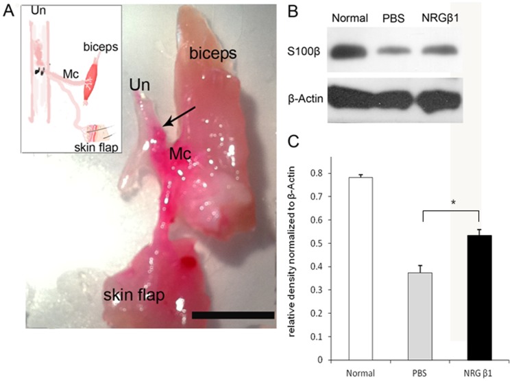
Figure 6. Photograph (A) and schematic diagram [inset in (A)] showing the effects of neuregulin β1 (NRG β1) on the process of nerve regeneration two months after end-to-side neurorraphy (ESN).
The regenerated axons were labeled by the retrograde tracer DiI (red). Note that after NRG β1 treatment, the majority of axons from ulnar nerve (Un) successfully innervates the biceps muscle and skin flap via the side-implanted musculocutaneous nerve (Mc, arrow indicates the suture site. Scale bar = 1 cm). Also note that this good neuro-regeneration was accompanied by the higher amounts of Schwann cells migrated to the Mc following NRG β1 as compared to that of phosphate buffer saline (PBS) treated group [detected by S100β immunoblotting (B) and expressed in quantitative histogram (C)]. *P<0.05 as compared to that of PBS treated value.

