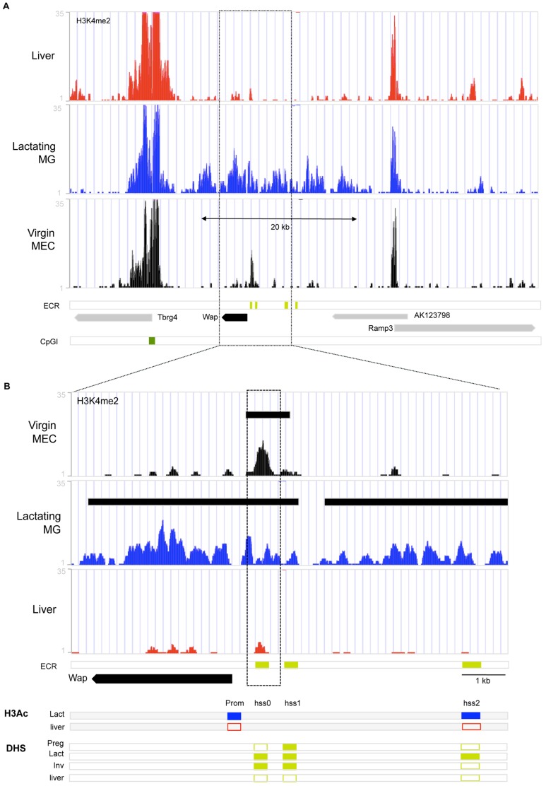
Figure 3. Chromatin organization at genomic region around Whey Acidic Protein gene (Wap).
Summary of markers of chromatin organization aligned to the Wap region in mouse genome assembly mm9 in the UCSC Genome Browser. (A–B) ChIP-seq reads for H3K4me2 in liver tissue (Liver, red), lactating mammary gland (Lactating MG, blue) and mammary epithelial cells isolated from 12 week virgin mammary glands (Virgin MEC, black). ECR: genomic locations of DHS conserved in mouse and rabbit [17], [54], CpG island is indicated by dark green bar. (B) Close-up of Wap region from (A). H3Ac: Summary of H3Ac-ChIP results for Wap promoter and HSS2 (see also Fig. S3) in lactating mammary gland (Lact, blue) and Liver tissue (liver, red): closed rectangle indicates enrichment of H3Ac at site, open rectangle indicates lack of enrichment at site. DHS: Summary of DHS results for rabbit from [17], [54]. Closed rectangle indicates presence of DHS, open rectangle indicates absence of DHS.

