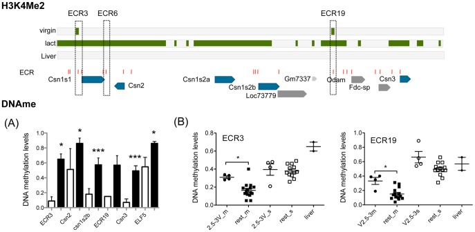
Figure 5. Tissue-specific DNA methylation, and DNA methylation for ECR3 and 19 during mammary gland development.
Top panel: Summary of H3K4me2 levels, ECR and gene locations in the CSN region (based on Figure 2). (A) Tissue specific DNA methylation levels: DNA methylation levels in lactating mammary gland (white bars) and liver tissue (black bars). Significance was determined using an un-paired, 2-tailed t-test: *<0.05, ***<0.0001. For ECR3 (MG, n = 2; Liver n = 2) and ECR19 (MG: n = 1; Liver n = 2) MeCpG percentages were determined with SEQUENOM mass-array technology (see table S2). Beta-casein promoter (Csn2) (MG, n = 5; Liver n = 5), Alpha-s2b casein promoter (Csn1s2b) (MG, n = 5; Liver n = 5), and Kappa casein promoter (Csn3) (MG, n = 4; Liver n = 4), were determined by bisulfite-sequencing (See Fig. 6 for details). (B) DNA methylation levels during mammary gland development and differentiation. MeCpG levels for ECR3 (average of 5 CpG sites in 561 bp) and ECR19 (average of 6 CpG sites in 599 bp) were determined with SEQUENOM mass-array technology (see table S2), different developmental time points are depicted with different symbols, each symbol represents an independent MEC or non-MEC prep from pooled tissue samples (see MEC prep) for virgin samples, 7day pregnant, INV and AMV, or individual animal (16 day pregnant and 8day lactating). Mammary epithelium (filled symbols) Pre-pubertal, filled circles (2.5-3V_m: 2.5–3 week old virgin females); Post-pubertal, filled square (rest_m: 3.5–4 week old virgins; 5.5–6 week old virgin females; 8–15 week old virgins; 7day pregnant females; 16 days pregnant; 8 day lactation; Inv (>28 day after lactation); Age Matched Virgin (Virgin animals same age as >28 day involuted animals). Non-MEC cell fraction of the mammary gland (open symbols): pre-pubertal: 2.5-3V_s, open circles: 2.5–3 week old virgin females; post-pubertal: rest_s (3.5–4 week old virgin females; 5.5–6 week old virgins; 8–15 Week old virgins; 7day pregnant females; Inv (>28 day after lactation); Age Matched Virgin (Virgin animals same age as >28 day involuted animals). Liver tissue : filled diamond.

