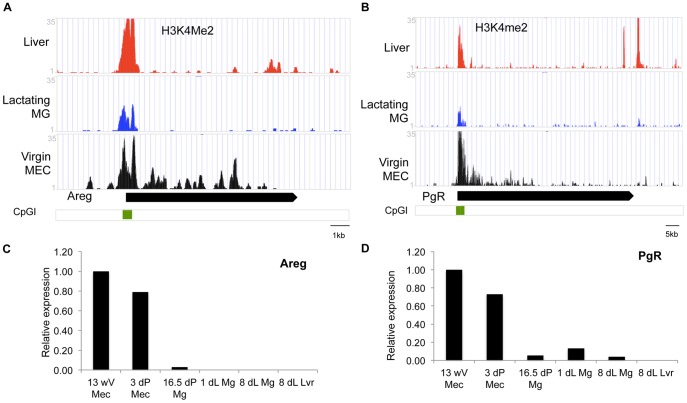
Figure 8. H3K4me2 ChIP-seq and RT-PCR results for genes expressed in virgin mammary gland.
(A–B): ChIP-seq reads for H3K4me2 in liver tissue (Liver, red), lactating mammary glands (Lactating MG, blue) and mammary epithelial cells isolated from 12 week virgin mammary glands (Virgin MG, black). (C–D): Q-RT-PCR of (C) Amphiregulin (Areg) and (D) Progesterone receptor PgR at different developmental stages in MEC isolated from mammary gland tissue of 13 week virgin (13wV), 3 days pregnant (3dP) or whole mammary tissue from 16.5 day pregnant (16.dP Mg) day 1 and day8 lactation (1dL Mg and 8dL MG) as well as liver from an 8d lactating animal (8dL Lvr). Real-time-RT-PCR data are normalized to Keratine 18 and expressed relative to 13wV MEC.

