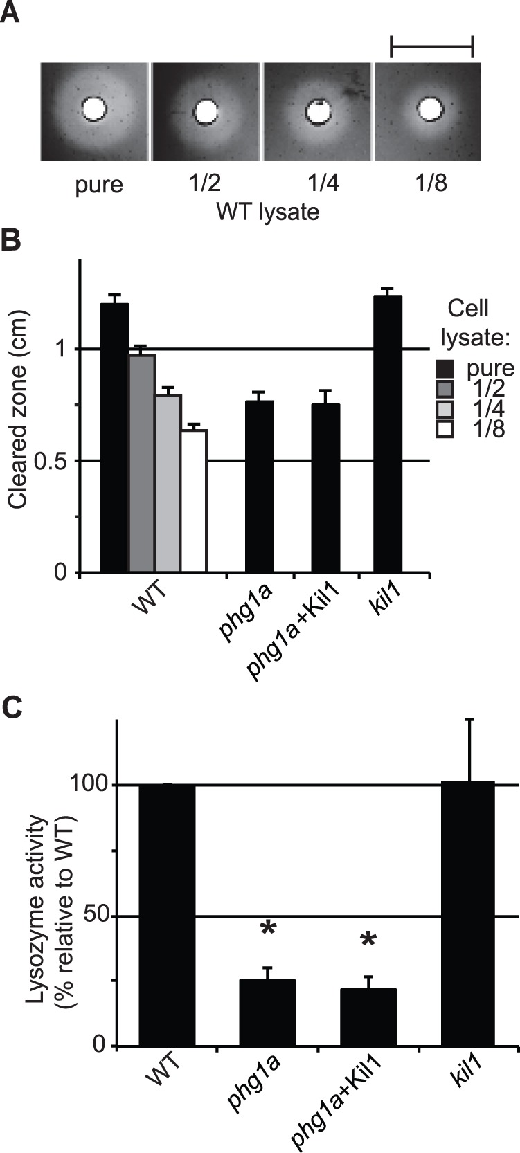
Figure 2. Cellular lysozyme activity is decreased in phg1a KO cells.

A) Lysate from WT cells were deposited on an agarose plate containing cell wall extracts of M. lysodeikticus. Lysozyme present in the lysate digests the bacterial cell wall, forming a cleared zone on the plate, the size of which decreased when the cellular lysate was diluted. Scale bar: 1cm. B) Average diameter of cleared zones for different concentrations of WT lysates provides a scale to which undiluted mutant cell lysates can be compared (n≥4) C) Lysozyme relative activity was deduced from results presented in B, after normalization. * : significantly different from WT (Student t-test; p<0.01).
