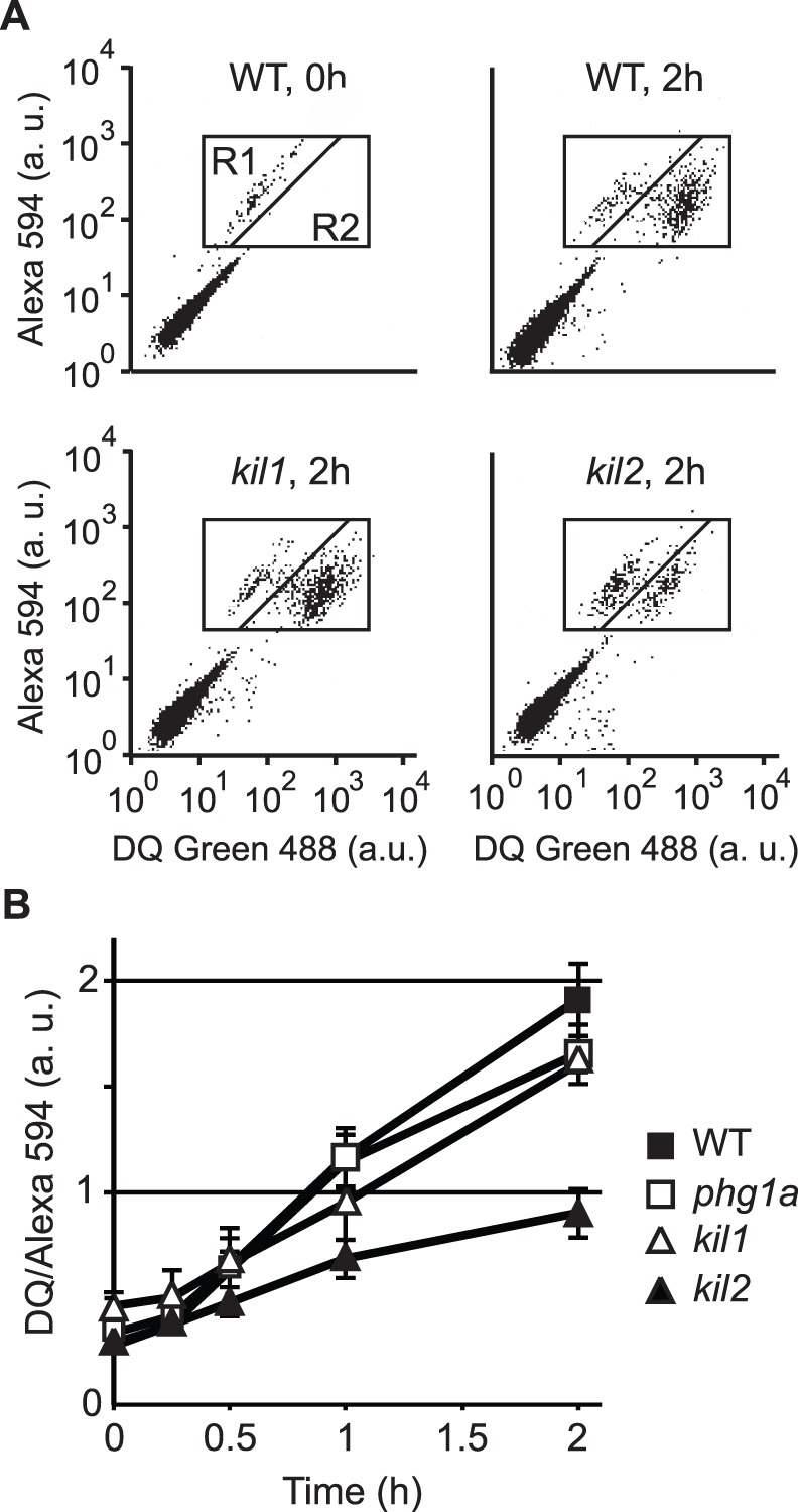
Figure 4. Phagosomal proteolysis is not defective in kil1 KO cells.

Cells were allowed to engulf silica beads coupled to Alexa Fluor 594 and to BSA labeled with DQgreen at a self-quenching concentration. Proteolysis of BSA in phagosomal compartments released DQgreen fluorescence, which was measured by flow cytometry in the R1+R2 region (cells containing fluorescent beads). A) WT cells having just phagocytosed beads (WT, 0 h) showed a low fluorescence (R1 window). After 2 h of incubation (WT, 2 h), the increase in DQgreen fluorescence revealed intra-phagosomal proteolytic activity. A similar pattern was observed in kil1 KO cells (kil1, 2 h), but not in kil2 KO cells (kil2, 2 h) where phagosomal proteolytic activity is reduced [8]. B) Cell-associated DQ green fluorescence was measured in the R1+R2 region after various times of incubation. The average and S.E.M. of 4 independent experiments are presented.
