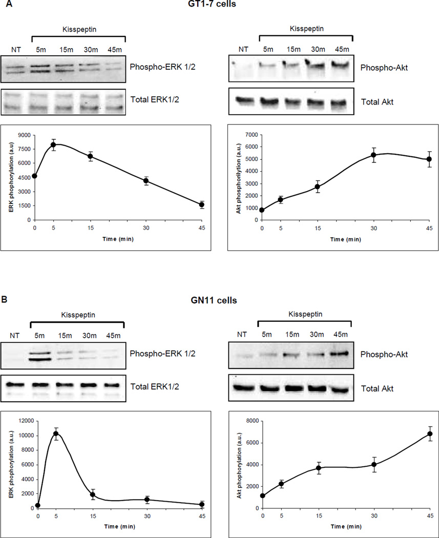
Figure 6. Expression and activation of Akt and p42/44 MAPK in GT1-7 and GN11 cells.
(A) GT1-7 and (B) GN11 cells. Western blots were performed in a time course over 45 min after incubation with 10−9M kisspeptin. Protein was analyzed for expression and activation of Akt using primary antibodies against phospho-Akt at Ser473 and p42/44 MAPK using primary antibodies against phospho-p44/42 MAPK at Thr202/Tyr204. Total levels of Akt and p42/44 MAPK were not altered by kisspeptin. The data were quantitated and the means±SE as arbitrary units (a.u.) are shown in the lower panels (n=3).

