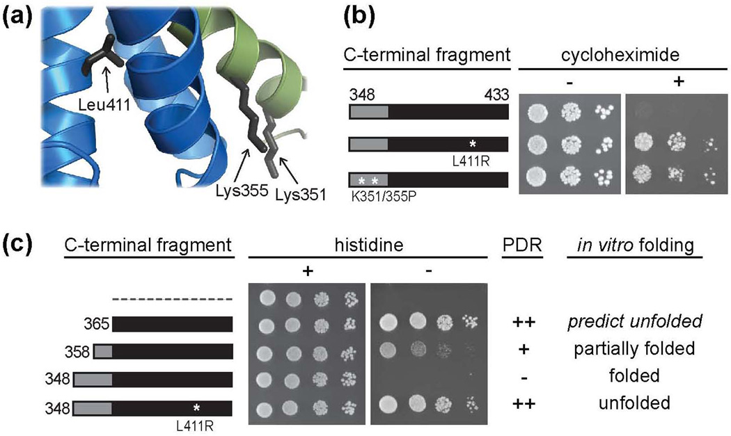
Figure 3.
Unfolding of Zuo1’s C-terminal helical bundle releases autoinhibition. (a) Ribbon diagram of a portion of the CTD with side chains of residues predicted to disrupt domain structure upon substitution indicated. (b) Wt cells were transformed with DNA encoding TAP tag fusions of the CTD with or without a destabilizing L411R or K351/355P alteration. Serial dilutions of cells containing the indicated plasmids were spotted onto medium without (−) or with (+) cycloheximide. (c) Modified yeast two-hybrid and correlation between protein fold and PDR induction. Cells containing an integrated Gal1-HIS3 reporter were transformed with DNA encoding the Gal4 DNA binding domain (GBD) alone (dotted line) or GBD fused to fragments of the C-terminus of Zuo1, with the N-terminal residue of each fragment indicated. Transformants were spotted in serial dilutions onto medium with (+) or without (−) histidine. Table includes summarized PDR induction data from Fig. 2a and Fig. 3b with (++) representing a high level of induction and (−) representing no induction and summarized in vitro folding data from Fig. 2c and Supplementary Fig. 2a. Zuo1365–433 is predicted to be unfolded (see text).

