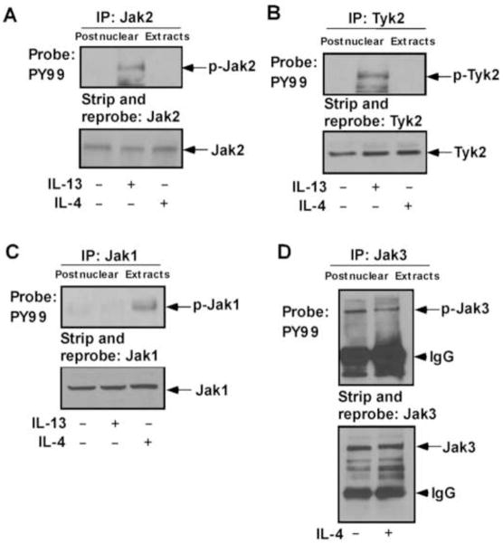
Figure 1. IL-4 and IL-13 activates different Jak kinases in monocytes.
Human blood monocytes (10×106/group) were directly stimulated with IL-13 or IL-4 for 10 min or left untreated as indicated. Cells were lysed and the postnuclear extracts were immunoprecipitated (IP) with different Jak/Tyk antibodies as mentioned in panels A–D. The immune complexes were analyzed by immunoblotting with antibody to phosphotyrosine, PY99 and presented in the upper panels of A–D. The bottom panels represent stripping and reprobing the blot with the same individual antibody used for immunoprecipitation, to assess equal protein loading. Arrows indicate the positions of respective Jak kinases as mentioned, based on the migration of molecular weight markers that were run in adjacent lanes. The arrowhead marks the migration of the heavy chain of IgG. Data are from a representative experiment of three independent experiments that were performed.

