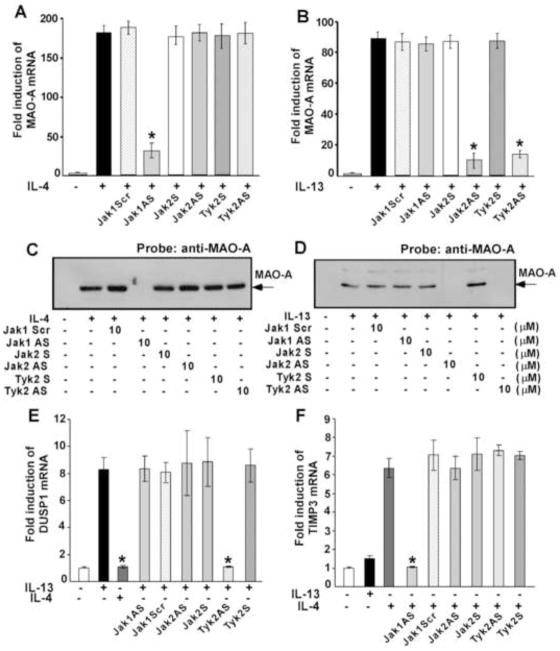
Figure 3. IL-4 and IL-13-stimulated MAO-A expression as well as IL-13 and IL-4 specific induction of DUSP1 and TIMP3 is controlled by different Jak kinases in human monocytes/macrophages.
Monocytes (5×10⁶/group) were pre-treated directly with antisense ODN to Jak1 or a scrambled ODN control (10 μM) (A–F) or Jak2 or Tyk2 antisense or sense ODNs (10 μM) (A–F) for 48 h, with one re-feed at 24 h, prior to the incubation with IL-4 (A, C, E, F) or IL-13 (B, D, E, F) for another 24h (A–D) or 48h (E, F). In panels A–B, total cellular RNA extracts were prepared and subjected to real-time quantitative PCR analysis. After normalization with GAPDH amplification, the fold induction of MAO-A mRNA expression for different groups was plotted. Data are the means ± S.D. (n=3). Significant differences were determined by comparing the antisense (AS), scrambled (Scr) and sense (S) ODN treated groups to the IL-4 or IL-13-treated control (*p<0.002) In panels C–D, cells were lysed and 50μg of postnuclear extracts were separated by SDS-PAGE and immunoblotted with an antibody against MAO-A. The data shown are representative of three separate experiments giving similar results.
For panels E and F, real-time PCR analyses was performed and fold induction of DUSP1 (E) and TIMP3 (F) mRNA expression for different groups was plotted after normalization with GAPDH. Data are the mean ± SD; (n=3). *p<0.003 compared to the IL-13 (E) or IL-4 (F)-treated controls.

