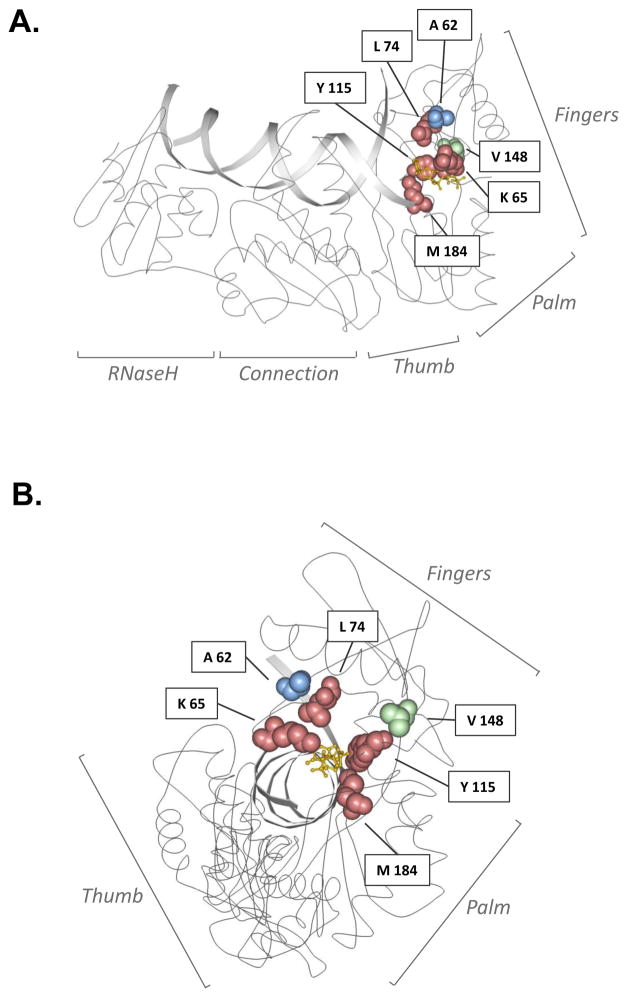
Figure 5. Locations of amino acid residues in HIV-1 reverse transcriptase (RT) associated with drug resistance and/or enzyme fidelity.
A ribbon diagram of HIV-1 RT is shown in side-view (A) or head-on (B) view of the catalytic domain, with interlaid primer:template DNAs, including a space-filling of amino acid residues investigated in this study. HIV-1 RT subdomains are indicated, and amino acid residues investigated in this study are represented as space-filled and are color-coded to indicate phenotype. Red denotes primary drug-resistant mutation site; blue denotes secondary drug-resistant mutation site; green denotes non-drug-resistant site. The yellow molecule depicts a dideoxynucleotide at the polymerase active site. Image adapted from of Huang, H. et al.96 and created with Protein Workshop software 97. Image obtained from the Research Collaboratory for Structural Bioinformatics (RCSB) Protein Data Bank (PDB) 98; IPDB ID: 1RTD.

