Figure 3.
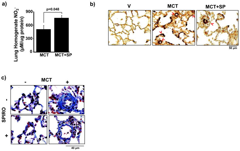
Spironolactone increases pulmonary vascular NO• levels and attenuates pulmonary vascular remodeling in PAH. (a) The effect of spironolactone (SP)(25 mg/kg/d) on pulmonary vascular NO• levels in PAH was assessed by measuring nitrite (NO2−) in lung tissue homogenates from Sprague-Dawley rats treated with vehicle control (V) or monocrotaline (MCT) (50 mg/kg) (n=4). (b) Tissue sections were stained with anti-smooth muscle cell α-actin antibody and the number of muscularized distal pulmonary arterioles (red arrows) was counted in 20 consecutive fields per section (100x magnification). Compared to V-treated rats with PAH, spironolactone decreased significantly the number of α-actin-stained muscularized distal pulmonary arterioles (76.0 [64–95] vs. 59.5 [59–61] muscularized pulmonary arterioles/20 high powered fields, p<0.005, n=5). (c) Gomori’s trichrome stain was performed on paraffin-embedded lung sections and perivascular collagen deposition in pulmonary arterioles measuring 20–50 μm located distal to terminal bronchioles (400x magnification) was measured. Compared to V-treated rats with PAH, spironolactone decreased perivascular collagen deposition by 77% (p<0.001, n=4–5 rats per condition). Representative photomicrographs are shown.

