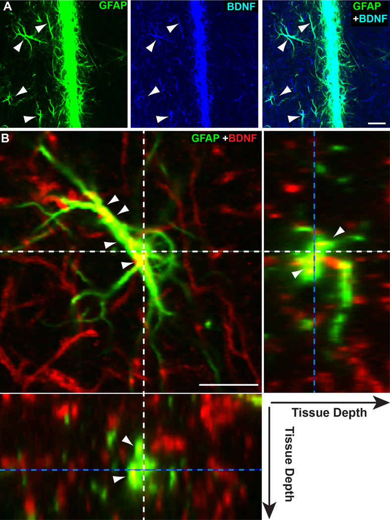
Figure 5. Localization of BDNF within astrocytes.
A) High magnification of single-plane (1.5 µm thick) through the midline between the two SC from the 10 mo DBA/2J shown in Figure 4C. Image shows apparent localization of BDNF within GFAP-labelled hypertrophic astrocyte processes (arrowheads). B) Single-plane (1.5 µm thick) image of a GFAP-labelled astrocyte near the dorsal ridge of an 8 mo DBA/2J SC (left panel). End-on (orthogonal) view of the same plane for both the Y (right panel) and X (left panel) axes illustrates location in tissue of the image plane (dotted blue lines) and of center of the astrocyte (dotted white lines). While most BDNF distributes in the underlying neuropil, localization within the astrocyte forms diffusely distributed pockets within the cytoplasm (arrowheads). Smaller astrocyte processes, possibly end-feet, that are associated with neuronal processes appear to contain far less BDNF. Scale = 20 µm (A) or 10 µm (B).

