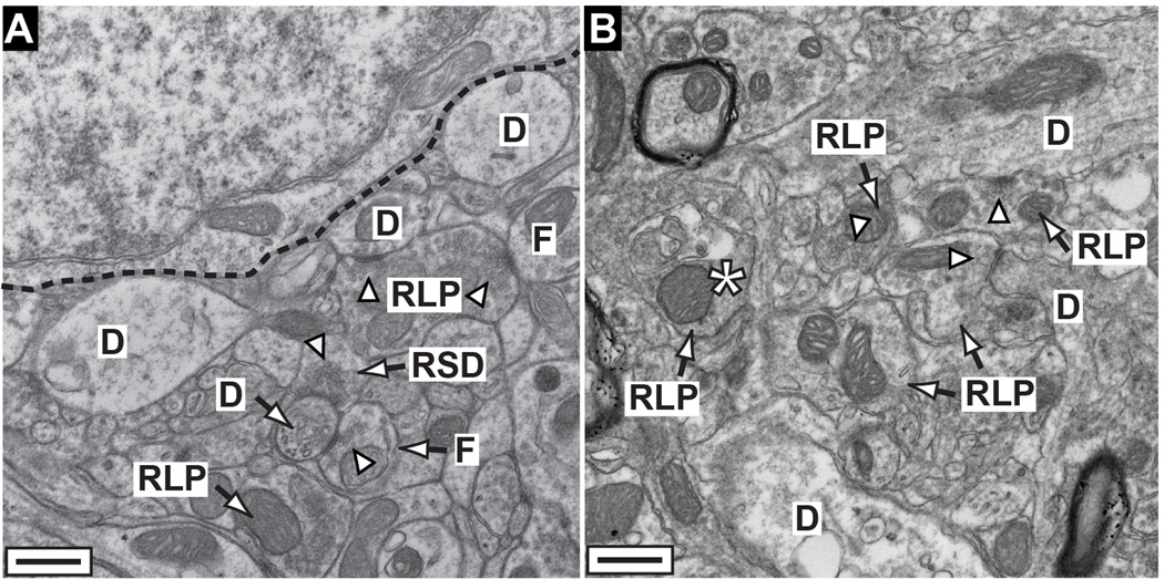
Figure 7. Structural persistence following transport depletion.
A) Electron micrograph of sagittal section through superficial SC from a 12 mo DBA/2J shows axon terminals from RGCs (RLP), intracollicular inhibitory neurons (F), and cortico-collicular projections (RSD) in proximity to a collicular relay neuron (dashed line) and dendrites (D). For definitions, see Calkins et al. (Calkins et al., 2005). Many terminals contain intact presynaptic active zones (arrowheads). B) A 15 mo DBA/2J colliculus also contains intact RLP terminals with some synapses to dendrites. Terminals near a degenerating RLP (*) appear slightly dystrophic.

