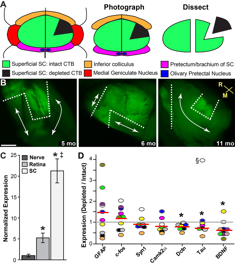
Figure 8. Gene expression with loss of transport in DBA/2J superior colliculus.
A) Schematic of a mouse brain in horizontal section showing orientation of the superficial superior colliculus (SC) in relation to other structures. The SC and surrounding tissue are isolated ex vivo, flat-mounted on a slide, and photographed. Finally, regions with intact CTB signal (green) and regions with depleted signal (black) are micro-dissected from each other and processed for RNA extraction. B) Representative fluorescent micrographs of three freshly-dissected and flattened DBA/2J SCs. For each, we isolated regions of intact (within dotted lines) and depleted (arrows) transport, followed by RNA extraction. Rostral and medial orientation indicated. Scale = 200 µm. C) Quantitative PCR measurements of Bdnf mRNA in retina, myelinated optic nerve, and super colliculus of 3 mo C57. For each of 5 animals, tissues from the left and right retinal projections were pooled. Data calculated relative to 18s rRNA and normalized to expression in the nerve (mean ± sd). Compared to Bdnf mRNA in the optic nerve, expression in retina was 5-fold more abundant and in SC 21-fold more abundant (*; p<0.001). SC was 4-fold greater than retina (‡; p<0.001). D) Quantitative PCR measurements of select genes in 10 individual SCs (circles) shown as ratio of expression in region of depleted to intact transport, with mean ratio for each gene indicated (red line). For each micro-dissected region, expression was performed in triplicate and normalized to 18s rRNA before calculating the ratio. Significance: * p=0.0004 (Dctn), p=0.017 (Tau), and p=0.008 (Bdnf) compared to hypothetical ratio of one (dotted line) using chi-square. Outlier excluded (§, p<0.001).

