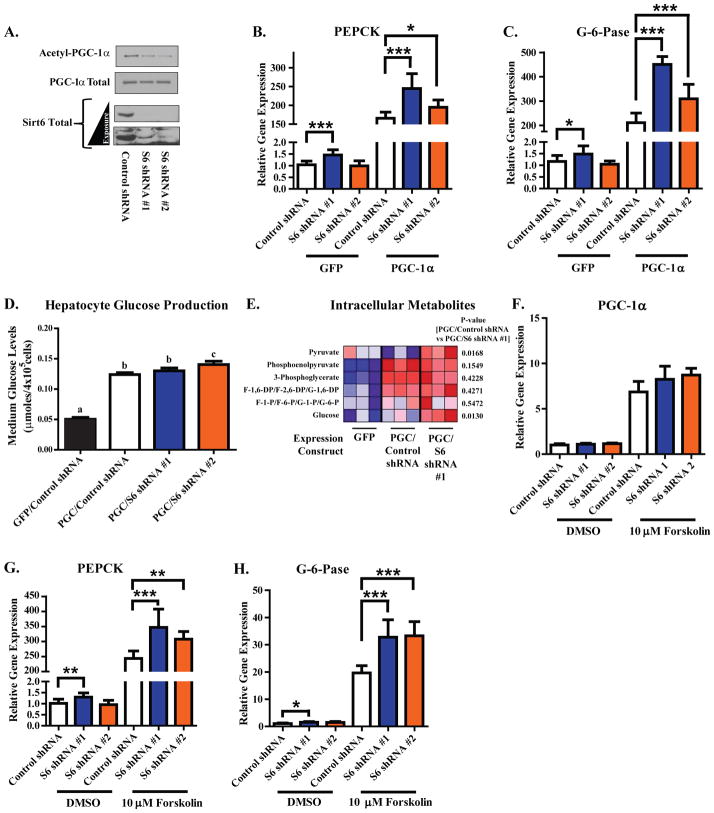
Figure 5. Reduction of endogenous Sirt6 decreases PGC-1α acetylation and enhances the gluconeogenic program of murine primary hepatocytes.
(A) Assessment of PGC-1α acetylation and Sirt6 protein levels in murine primary hepatoctyes infected with the indicated adenoviral shRNA-expression constructs. (B,C) qRT-PCR measurement of PEPCK (B) and G-6-Pase (C) mRNA levels in primary hepatoctyes infected with GFP or PGC-1α and the indicated shRNA constructs. Data in Figures (A–C) are from three independent experiments. (D) Glucose production in primary hepatocytes infected with either GFP or PGC-1α and the indicated shRNA constructs. Data are pooled from two independent experiments. Columns with different letters above them are statistically significant (p<0.05). (E) Heat map depicting LC-MS analysis of intracellular metabolites from primary hepatocytes expressing the indicated constructs. (F–H) Measurement of PGC-1α (F), PEPCK (G), and G-6-Pase (H) mRNA levels in primary hepatoctyes infected with the indicated shRNA expression constructs and treated for 1.5 h with DMSO vehicle or 10 μM forskolin. In Figures (F–H), data are pooled from three independent experiments. All data expressed as means±S.E.M. *-p<0.05; **-p<0.01; ***-p<0.001 by one-way ANOVA with a Tukey’s post-test. See Figure S4.

