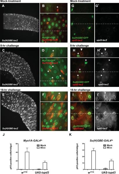
Fig. 3. upd3 is expressed and required in differentiated ECs and in differentiating EBs.
(A, C and E) Confocal microscopy images of posterior midgut (10× objective, XY section). (B, D, F, G, H and I) Confocal microscopy images of posterior midgut (60× objective). Bottom panels show XY section. Dotted lines indicate Y coordinates for XZ reconstruction (top panels).
(A–F) Expression of the EC-specific marker Myo1A and EB-specific marker Su(H)-lacZ in mock-treated animal (A and B), animals infected for 8 hours with Ecc15 (C and D) and animals infected for 18 hours with Ecc15 (E and F).
(G–I) Expression of the EB-specific marker Su(H)>GFP and the upd3-lacZ reporter in mock-treated animal (G), animals infected for 8 hours with Ecc15 (H) and animals infected for 18 hours with Ecc15 (I). (J and K) pH3-positive nuclei were scored in Myo1A-Gal4ts/+ (w1118) or Myo1A-Gal4ts/UAS-iupd3 (UAS-iupd3) (J) and Su(H)-GAL4ts/+ (w1118) or Su(H)-Gal4ts/UAS-iupd3 (UAS-iupd3) (K) animals infected with Ecc15 for 8 hours. Averages of three representative experiments (n = 12 guts/experiments) with s.d. are shown. (J) UAS-iupd3 vs. w1118, P = 0.0462 (*). (K) UAS-iupd3 vs. w1118, P = 0.0164 (*).

