
Fig. 5. The Upd3 cytokine produced in EB locates to the extracellular matrix and activates JAK/STAT signaling in EB and in visceral muscles.
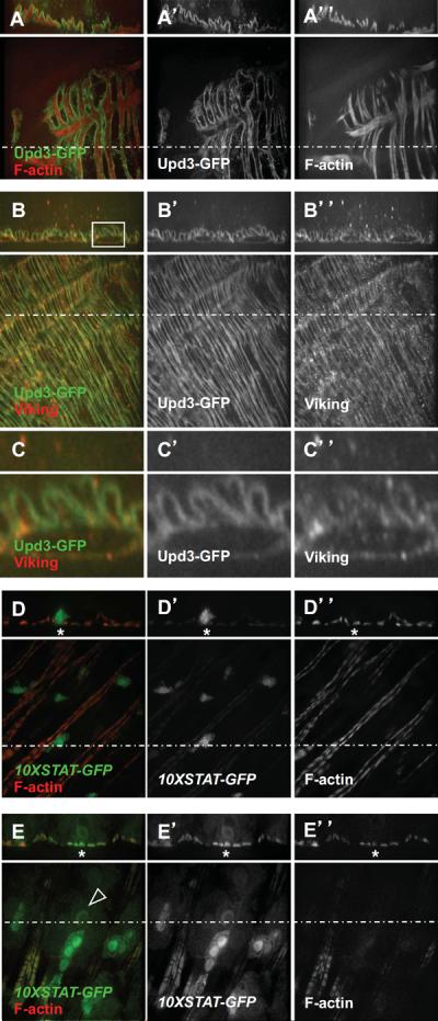
(A) The Upd3-GFP fusion decorates muscles as visualized by F-actin staining.
(B) The Upd3-GFP fusion protein co-localizes with the extracellular matrix marker Viking.
(C) Magnification of the squared area shown in (B).
(D) The 10X-STAT-GFP reporter is expressed in EBs and display basal expression level in visceral muscles.
(E) Over-expression of the UAS-upd3 transgene using the Su(H)-Gal4ts driver leads to 10X-STAT-GFP expression in visceral muscles (asteriks) and differentiation of EBs (arrowhead pointing to the nucleus of a differentiating EBs).